


PTT-6®️ Stimulates Elastin and Hyaluronic Acid Production in Eyelid Skin Leading to Improved Aesthetics
Published in the peer-reviewed journal Dermatologic Surgery, this article studies the pathways in which red deer umbilical cord lining conditioned media (PTT-6®️) stimulates fibroblast elastin and hyaluronic acid production to improve the look of the eye area; includes observation of users with photographic comparison demonstrating subjective and objective improvement.

As seen in
![]()
BACKGROUND
Conditioned media (CM) from red deer umbilical cord lining mesenchymal stem cell (RD-CLMSC) culture comprises an exosomal suspension and is the active ingredient of a facial cream (CALECIM Professional Multi-Action Cream, CALECIM Cosmeceuticals, Singapore) shown to significantly improve skin tone after topical application. This effect appears to be related to increased human dermal fibroblast (HDF) proliferation, with increased HDF production of elastin and hyaluronic acid (HA).
OBJECTIVE
The authors sought to investigate if RD-CLMSC-CM might increase elastin and HA to improve thin eyelid skin turgor and elasticity.
MATERIALS AND METHODS Upper eyelid skin HDF from 9 individuals were exposed in culture to RD-CLMSC-CM. Photocolorimetry after histochemical staining demonstrated increased expression of elastin by 68% and HA by 113% compared with exposure to Dulbecco’s modified eagle medium/10% fetal calf serum. A topical eyelid skin formulation containing RD-CLMSC-CM was applied to the upper and lower eyelid skin of 4 preexisting users of RD-CLMSC-CM facial cream. Photographic comparison before and after application demonstrated subjective and objective cosmetic improvement.
RESULTS AND CONCLUSIONS
Topical application of RD-CLMSC-CM increased eyelid HDF production of elastin and HA. This improved skin elasticity and turgor to heighten eyelid skin aesthetics

Cord Lining Epithelial Stem Cells (CLEpSC) and Cord Lining Mesenchymal Stem Cells (CLMSC) were first isolated from the outer lining of the umbilical cord in 2004. It quickly became apparent that this was an economically viable source of stem cells as an enormous number of cells could be harvested up to passage 30 with no change in karyotype, phenotype, or stemness. In multiple case series, Human Cord Lining Mesenchymal Stem Cells (H-CLMSC) were found to efficiently heal a variety of wounds when either applied directly to the wound or when seeded on Biobrane (Smith and Nephew, Hull, United Kingdom) and applied on the wound. With the understanding that intercellular communication is accomplished by paracrine signaling, sterile filtered conditioned media (CM) derived from CLMSC culture was collected and found to also accelerate wound healing in multiple case series (Author’s unpublished Information).
After rigorous preclinical studies, a United States Food and Drug Administration (USFDA) Investigational New Drug (IND) trial using Current Good Manufacturing Practice (cGMP) grade allogeneic H-CLMSC (Biological drug name Corlicyte®) to heal chronic diabetic foot ulcers was commenced in Colorado USA. In phase 1, 9 subjects in 2 dosing cohorts completed the trial. No subjects experienced a serious adverse reaction to Corlicyte® or the development of anti–human leukocyte antigen (HLA) antibodies. Sixty percent of subjects in the lower dose cohort experienced ulcer closure by Day 70 of follow-up, whereas the mean ulcer size was reduced by 54% to 67% in the other subjects. Phase 2 of this trial will commence in due course.
Mesenchymal Stem Cells (MSCs) secrete extracellular vesicles (EVs) to exert paracrine effects on cells, which internalize the secreted EVs. EVs are a heterogenous collection of membrane-bound carriers including exosomes, which contain complex cargos including protein cytokines and growth factors, lipids, and nucleic acids. These contents contribute to the varied components of CM, which have been shown to promote tissue repair, stimulate hair follicle regeneration, 10 and accelerate wound healing. 11,12 A list of protein cytokines and growth factors found in H-CLMSC-CM is found in Table 1.
A cosmeceutical line based on CLMSC-CM was developed closely following directives from Cosing, the official cosmetic ingredient database of the European Union as well as the Association of South East Asian Nations Cosmetic Directive, both of which prohibit cosmetic products containing human derivatives. Red deer (RD) were selected as the donor animal for umbilical cord outer lining membrane as these animals do not harbor transmissible prion disease such as bovine spongiform encephalopathy and are ethically farmed for horn velvet in New Zealand. RD-derived products are approved for use and listed in Cosing.
In a double blinded, randomized controlled trial, application of RD-CLMSC-CM as a serum or as a cream resulted in decreased erythema and crusting after ablative laser treatments with improvement in wrinkling. Facial rejuvenation was also observed after regular topical application of RD-CLMSC-CM to the skin in a transepidermal vehicle. Red deer umbilical cord lining mesenchymal stem cell–conditioned media in a gel formulation applied to the periwound skin was additionally shown to accelerate chronic wound healing.
In vitro, RD-CLMSC-CM was demonstrated to be as effective as H-CLMSC-CM in stimulating human dermal fibroblast (HDF) proliferation as well as in increasing HDF elastin and hyaluronic acid (HA) production demonstrating mammalian cross-species efficacy.
In this study, the authors sought to investigate how RDCLMSC-CM would alter the in vitro expression of HA and elastin in cultured HDF from eyelid skin. Four preexisting RD-CLMSC-CM facial cream users were also recruited for a volunteer case series to assess the clinical effects of RDCLMSC-CM application on eyelid skin after a 35-day period with photodocumentation before and after treatment to assess clinical results.

Materials and Methods
Red Deer Umbilical Cords From New Zealand
Red deer umbilical cords were collected under specific handling instructions from a farm in New Zealand that rears RD for the collection of horn velvet. The cords were transported in DMEM/penicillin/streptomycin/amphotericin B between 2° and 8°C for the entire transfer duration from the farm to the laboratory in Singapore.
Isolation of Red Deer Cord Lining Mesenchymal Stem Cell
Red deer cord lining mesenchymal stem cells were isolated from umbilical cords according to the protocol described in international patent publication number WO 2006/ 019,357 A1. Briefly, the umbilical cords were washed in PBS supplemented with penicillin/streptomycin/amphotericin B until the solution turned clear. The amniotic membrane was divided into small pieces, placed on the plastic surface of the culture flask, submerged in PTT-6 medium (CellResearch Corp, Singapore), and incubated at 37°C with 5% CO 2 . The culture medium was changed every 2 to 3 days. At a confluence of about 70%, cells were trypsinized (0.05% trypsin/0.02% ethylenediaminetetraacetic acid) for further expansion or for cryo-preservation.
Eyelid Skin–Derived Human Dermal Fibroblasts
Human dermal fibroblasts were isolated from eyelid skin of donors with varying profiles, as detailed in Table 2. Samples were obtained from the tissue bank of CellResearch Corporation, a registered tissue bank under the Ministry of Health, Singapore. The tissue bank operates in compliance with the Human Tissue Framework created under the Human Biomedical Research Act (2015).

Derivation of Conditioned Media From Red Deer Cord Lining Mesenchymal Stem Cell Culture
Cryovials containing RD-CLMSC were retrieved from storage and quick thawed in a 37°C water bath. PTT-6 medium (CellResearch Corporation, Singapore) was used for culturing CLMSC (RD & H) at 37°C with 5% CO 2 . PTT-6 was changed every 2 to 3 days. At 100% confluency, PTT-6 was replaced with DMEM basal medium. Culture dishes were incubated for 48 hours, and spent medium was collected into centrifuge tubes for centrifugation at 1800 rpm for 10 minutes. The supernatant was collected as conditioned medium (CM) into labelled tubes: RDCLMSC-CM and kept at 280°C until use.
Hyaluronic Acid and Elastin Staining
Eyelid skin–derived HDFs (refer to Table 2) were seeded into 96-well plates at a density of 10,000 cells per well in DMEM/10% fetal calf serum (FCS) (Life Technologies, Cat. No. 10270106). Once cells reached approximately 80% confluence, the medium was removed and cells were washed once with phosphate-buffered saline (PBS). They were then cultured in either DMEM/10% FCS (control) or RD-CLMSC-CM for 48 hours.
After treatment, immunocytochemical staining was performed using anti-HA (MyBioSource, Cat. No. MBS2025717) and antielastin (Life Technologies, Cat. No. MA127129) antibodies. Briefly, cells were washed with PBS, fixed in cold methanol for 10 minutes, and blocked with 2.5% normal horse serum (Vector Laboratories, Cat. No. PK-7200) for 20 minutes in a humidified chamber. Cells were then incubated with primary antibodies for HA (1:50) and elastin (1:50) for 2 hours at room temperature.
After incubation, cells were washed with Tris-buffered saline (TBS; VWR Life Science, Cat. No. 788) containing 1% Tween-20 (Sigma, Cat. No. P9416), followed by a 15minute incubation with secondary antibodies (Vector Laboratories, Cat. No. PK-7200). This was followed by 3 additional washes with TBS/1% Tween solution. Cells were then treated with ABC reagent (Vector Laboratories, Cat. No. PK-7200) for 15 minutes at room temperature.
Colorimetric development was performed using DAB substrate (Dako, Cat. No. K3468), and Harris hematoxylin (Sigma, Cat. No. HHS16) was used for nuclear counterstaining. Images were captured using a bright-field phase contrast microscope (Olympus) at 103 magnification. Quantification of HA and elastin expression was conducted using ImageJ software (National Institutes of Health, USA).
Clinical Improvement in Eyelid Skin Reduction of Fine Lines and Wrinkles
Having established the nontoxicity and efficacy of RDCLMSC-CM in a transepidermal vehicle (Lynk Biotech, Singapore) for facial skin rejuvenation from an earlier study, 4 preexisting users of CALECIM Professional skin care products volunteered to use the eye cream (CALECIM Professional Eye Contour Lifting Cream, CALECIM Cosmeceuticals, Singapore) twice daily for 5 weeks with photographs taken at baseline and after the trial period for clinical assessment using a global aesthetic improvement scale (GAIS) for both the clinician and the patient. Evaluated variables included rhytids, skin texture, laxity, dyschromia, and the appearance of the eyebags. Subjects rated their improvement with a self-assessment questionnaire. Clinician GAIS was based on a 5-point scale (1—much improved, 2—improved, 3—no change, 4—worse, 5—much worse). No major or minor adverse effects were reported throughout the study period.
Results
Effect of Red Deer Umbilical Cord Lining Mesenchymal Stem Cell–Conditioned Media on the Expression of Human Dermal Fibroblast Elastin and Hyaluronic Acid
To evaluate the effect of RD-CLMSC-CM versus DMEM/ 10% FCS (control) on HA and elastin expression in eyelid skin–derived HDFs, cells from 9 donors aged 23 to 69 years were selected from CRC’s skin cell library. Human dermal fibroblasts were cultured for 48 hours in either DMEM/ 10% FCS (control) or RD-CLMSC-CM, followed by immunocytochemical staining for HA and elastin.
Expression levels were quantified as optical densities using ImageJ (NIH). Human dermal fibroblasts exposed to RD-CLMSC-CM showed a 113% increase in HA expression and a 68% increase in elastin expression compared to DMEM/10% FCS (control) (Figure 1). These effects were consistent across all donor age groups, suggesting that the upregulation of HA and elastin by RD-CLMSC-CM is independent of donor age.
 Figure 1. Representative immunocytochemical images showing HA and elastin expression in HDFs cultured for 48 hours in either DMEM/10% FCS (control) or RD-CLMSC-CM. Human dermal fibroblasts (n 5 9), derived from eyelid skin of donors aged 23 to 69 years, were stained for elastin and HA. Quantification of optical density using ImageJ revealed that RD-CLMSC-CM treatment increased HA and elastin expression by 113% and 68%, respectively, compared to DMEM/10% FCS (control). DMEM, Dulbecco’s modified eagle medium; FCS, fetal calf serum; HA, hyaluronic acid; HDF, human dermal fibroblast; RD-CLMSC-CM, red deer umbilical cord lining mesenchymal stem cell–conditioned media.
Clinical Improvement in Eyelid Skin Reduction of Fine Lines and Wrinkles
Four existing RD-CLMSC-CM cream (CALECIM Professional Multi Action Cream, CALECIM Cosmeceuticals, Singapore) users volunteered to try RD-CLMSC-CM in a formulation suitable for the eyes (CALECIM Professional Eye Contour Lifting Cream, CALECIM Cosmeceuticals, Singapore). Pretreatment photographs were taken, after which the volunteers were advised to apply the RDCLMSC-CM eye cream twice a day after cleansing to both upper and lower eyelids. They returned after 35 days (5 weeks) to complete the self-assessment questionnaire and have their photographs taken for objective assessment by a medical professional.
Overall, all 4 volunteers in this small series self-reported improvements in laxity and eye bag appearance. Approximately 75% reported improvements in rhytids, texture/fine lines, and dyschromia. Investigator assessment showed improvements in all volunteers for texture/fine lines, laxity, and eye bag appearance, with 75% improvement in rhytids and 50% improvement in dyschromia (Table 3; Figures 2–5).
<
Discussion
Of all the facial structures of the face that affect the perception of age, the periorbital receives the most attention. Typical morphologic characteristics influencing perceived age include crows’ feet wrinkles, undereye wrinkles, fine lines in both upper and lower eyelids, eyebags, dark circles under the eyes, the degree of eye-opening, blepharoptosis, and nonpathologic drooping of the upper eyelid.
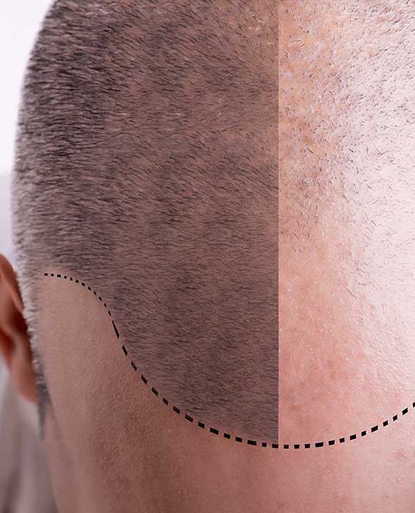
Less than 1 mm in thickness, eyelid skin is the thinnest skin in the human body and especially vulnerable to external insult in the form of photoaging from UV exposure as well as environmental irritants and pollutants. The overall thinness of eyelid skin with a thinner stratum corneum also offers lower impedance and higher drug permeation through eyelid skin regardless of drug lipophilicity. Anatomically, eyelid skin is in close apposition to the underlying orbicularis oculi muscle with no intervening subcutaneous fat.
Eyelid skin aging results from changes that are both intrinsic and extrinsic. Intrinsic skin aging is influenced by hormonal changes that occur with aging and deficiencies in estrogens and androgens. The net result, both in the dermis and epidermis, is tissue atrophy, skin wrinkling, dryness, and loss of elasticity of the skin. The most profound changes take place in the extracellular matrix (ECM), with decreases in the production of collagen, glycosaminoglycans like HA, and elastin.
Extrinsic aging is the result of exposure to external factors, largely UV irradiation and is also called photoaging. In UV exposed areas as the eyelids, the aging process is accelerated by superimposition of extrinsic skin aging on intrinsic skin aging.
Elastic fibers are composed of elastin and fibrillin microfibrils and are another major component of the ECM of skin, complementing the tensile strength of fibrillar collagens for this dynamic organ.
Degradation of the elastic fiber network is related to upregulation of elastic fiber degrading enzymes as MMP-2, -3, -9, -12, and -13, similar to that of collagen degradation, as well as serine protease neutrophil elastase.
Hyaluronic acid is a glycosaminoglycan and one of its most important properties is its strong hydrophilicity and ability to increase their volume in an aqueous environment. HA’s high osmotic activity and anionic structure enables it to bind large amounts of water. Over 50% of the total HA content is found in the skin and in human skin, HA is the key substance that fills the ECM with a key role to maintain optimal hydration and structural integrity with an important role in wound healing.
 Figure 2. Subject 2: Before (left) and after (right) 5 weeks of applying RD-CLMSC-CM eye cream. RD-CLMSC-CM, red deer umbilical cord lining mesenchymal stem cell–conditioned media.
 Figure 3. Subject 2: before (left) and after (right) 5 weeks of applying RD-CLMSC-CM eye cream. RD-CLMSC-CM, red deer umbilical cord lining mesenchymal stem cell–conditioned media.
As it is for collagen and elastin, intrinsic and extrinsic factors give rise to significant decrease in HA in the skin with aging, and the resultant loss of skin hydration results in skin wrinkles and sagging as well as impaired wound healing.
Conditioned media (CM) from RD-CLMSC culture comprises an exosomal suspension containing protein cytokines and growth factors in specific ratios and proportions and is the active ingredient of a facial cream (CALECIM Professional Multi-Action Cream, CALECIM Cosmeceuticals, Singapore), which has been shown to have positive effects on facial skin rejuvenation.
Topical application in the earlier studies did not include eyelid skin, and the authors sought to see its effects on this part of the face with a significant bearing on aging. To this end, an eye cream was formulated containing RD-CLMSCCM (CALECIM Eye Contour Lifting Cream, CALECIM Cosmeceuticals, Singapore) for topical application to the eyelids.
This study also sought to complement an earlier study 40 to investigate changes in HA and elastin production in eyelid HDF exposed to RD-CLMSC-CM and to correlate these changes clinically by examining photographs taken before and after treatment. Human dermal fibroblast isolated from upper eyelid skin of 9 donors of both sexes with ages ranging from 23 to 69 were subjected to 48 hours of culture in DMEM/10% FCS (control group), and RDCLMSC-CM. Optical density measurements of immunohistochemical staining revealed that RD-CLMSC-CM upregulated the expression of elastin by 68% and HA by 113% compared with the negative control.
Photographic evaluation of the eyelids of 4 volunteers in a case series before and after 5 weeks of topical application of RD-CLMSC-CM showed subjective and objective improvements in rhytids, texture/fine lines, elasticity, dyschromia, and the appearance of the eye bags. Visible results in a short time may likely be due to the thinness of eyelid skin and its attendant good absorptive characteristics.
These results suggest that RD-CLMSC-CM enhances ECM protein expression in eyelid-derived HDFs, supporting its potential use as a cosmetic ingredient for improving skin hydration and elasticity, regardless of donor age.
 Figure 4. Subject 3: before (left) and after (right) 5 weeks of applying RD-CLMSC-CM eye cream. RD-CLMSC-CM, red deer umbilical cord lining mesenchymal stem cell–conditioned media.
 Figure 5. Subject 3: before (left) and after (right) 5 weeks of applying RD-CLMSC-CM eye cream. RD-CLMSC-CM, red deer umbilical cord lining mesenchymal stem cell–conditioned media.
Conclusion
Red deer umbilical cord lining mesenchymal stem cell–conditioned media has now been shown to have rejuvenating effects on facial skin and the eyelids. This perceived clinical improvement in the eyelids is correlated to increased production of elastin and hyaluronic acid by eyelid HDFs. Further mechanisms at work may involve the exosomal creation of environments supportive of native stem cell populations in the skin.